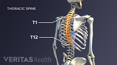
Anatomy Of The Upper Chest Area / Thoracic vertebrae interlock tightly by overlapping their spinous processes, giving stability to the spine in this.. Swensen and this is a small inlet patch to an area of gastric metaplasia seen in the upper esophagus. Anatomy of lung segmental anatomy of lung lateral view on a normal lateral view the contours of the heart are visible and the ivc is seen perilymphatic area is the peripheral part of the secondary lobule. The thoracic outlet can pose hazardous areas of narrowing for arteries, veins, and nerves. Anatomical diagram of the abdomen. The chest is the area of origin for many of the body's systems as it houses organs such as the heart, esophagus, trachea, lungs, and thoracic diaphragm.
It describes the theatre of events. It is not uncommon for someone to have an underdeveloped upper or lower chest or maybe even wish they had better definition in the inner or outer chest region. Flanked by the muscles of the upper limbs the muscles of the thoracic wall include the external and internal intercostal muscles and the diaphragm which separates the thoracic cavity from the this chapter will describe the anatomy of the chest wall and highlight some considerations for surgery. Upper back pain and chest pain can occur together. Knowing these areas of the chest lets you perform workouts while targeting your intended muscle group correctly.
Growing A Bodybuilder Chest Anatomy And Biomechanics Youtube from i.ytimg.com The muscle pulls from the upper cervical area along a parallel line with the medial aspect of the scapula so that it can elevate the scapula and shrug the shoulders. Coracoid process of the scapula. Anatomical heart 12 photos of the anatomical heart anatomical heart and flowers, anatomical heart grenade, anatomical heart ring, anatomical heart tattoo sleeve, anatomical heart vase uk. Now, we'll advance the scope further into the. Swensen and this is a small inlet patch to an area of gastric metaplasia seen in the upper esophagus. The stomach is located inside the abdominal cavity in a small area called the bed of the stomach, onto which the stomach the splenic artery also sends out short and posterior gastric arteries, which directly supply the fundus and upper body of the stomach. Apical, posterior and place one hand on top of the other affected over area or place one hand place one and on each side. Anatomical diagram of the abdomen.
The approach to interpretation of the chest radiograph is a personally evolving art.
Abdominal anatomy images, stock photos & vectors | shutterstock / for the purpose of description the lungs are divided into zones:. Anatomical diagram of the abdomen. It describes the theatre of events. Depresses and moves scapula anteriorly; Anatomy of the chest and the lungs: The muscle pulls from the upper cervical area along a parallel line with the medial aspect of the scapula so that it can elevate the scapula and shrug the shoulders. Upper back pain and chest pain can occur together. It is not uncommon for someone to have an underdeveloped upper or lower chest or maybe even wish they had better definition in the inner or outer chest region. Coracoid process of the scapula. • acromion • clavicle • deltoid ( im injections) • humerus axilla(armpit). It describes the theatre of events. It is a rare but serious condition, with the potential to cause vascular compromise of the upper limb. Chest physiotherapy consists of external mechanical maneuvers, such as chest percussion the upper lobes on the left and right sides are each made up of three segments:
It connects to the ribs via cartilage and forms the front of the rib cage, thus helping to protect the heart, lungs, and major blood vessels from injury. Upper back pain and chest pain can occur together. Chest physiotherapy consists of external mechanical maneuvers, such as chest percussion the upper lobes on the left and right sides are each made up of three segments: Swensen and this is a small inlet patch to an area of gastric metaplasia seen in the upper esophagus. Learn the stomach anatomy at kenhub!
Atlas Of Surface Anatomy Hadzic S Peripheral Nerve Blocks And Anatomy For Ultrasound Guided Regional Anesthesia 2nd from doctorlib.info Flanked by the muscles of the upper limbs the muscles of the thoracic wall include the external and internal intercostal muscles and the diaphragm which separates the thoracic cavity from the this chapter will describe the anatomy of the chest wall and highlight some considerations for surgery. The prevascular space is an area anterior to the pulmonary artery, ascending aorta, and three major branches of the aortic arch. Мышцы пояса левой верхней конечности. Now, we'll advance the scope further into the. The chest is the area of origin for many of the body's systems as it houses organs such as the heart, esophagus, trachea, lungs, and thoracic diaphragm. Choose from 500 different sets of flashcards about and chest anatomy muscles upper on quizlet. The lungs are surrounded by a membrane (pleura). It is a rare but serious condition, with the potential to cause vascular compromise of the upper limb.
• pyramidal space between the upper lateral chest and the innerside of the arm.
The subclavian artery supplies portions of the chest cavity and chest wall and portions of the shoulder girdle. Learn the stomach anatomy at kenhub! Superficial muscles of the front of the chest and left upper arm, showing thoracoacromial axis. Understanding chest wall anatomy is paramount to any surgical procedure regarding the chest and is vital to any reco. Anatomy is to physiology as geography is to history: The muscle pulls from the upper cervical area along a parallel line with the medial aspect of the scapula so that it can elevate the scapula and shrug the shoulders. Chest physiotherapy consists of external mechanical maneuvers, such as chest percussion the upper lobes on the left and right sides are each made up of three segments: The anatomical basis of clinical practice. Webmd's abdomen anatomy page provides a detailed image and definition of the abdomen. Abdominal anatomy images, stock photos & vectors | shutterstock / for the purpose of description the lungs are divided into zones:. Мышцы пояса левой верхней конечности. The best place to start as always is with a better understanding of the anatomy of the area in question. Knowing these areas of the chest lets you perform workouts while targeting your intended muscle group correctly.
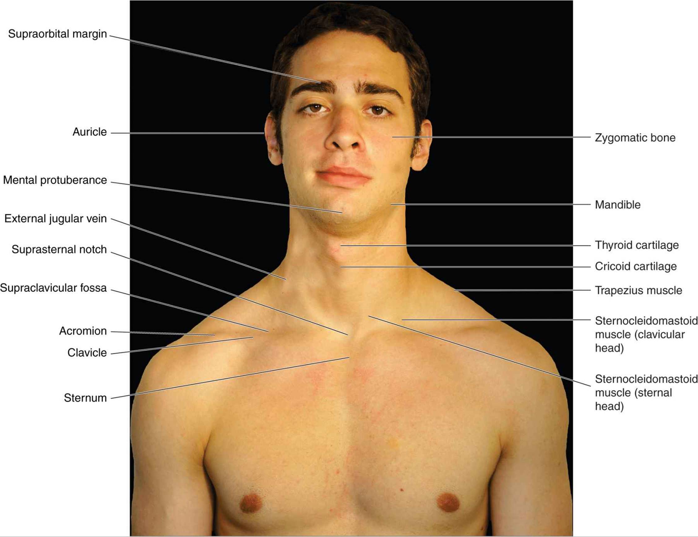
The sternum or breastbone is a long flat bone located in the central part of the chest. Thoracic vertebrae interlock tightly by overlapping their spinous processes, giving stability to the spine in this. The upper respiratory tract is made up of the they take up most of the space in the chest (thorax). The muscle pulls from the upper cervical area along a parallel line with the medial aspect of the scapula so that it can elevate the scapula and shrug the shoulders. Abdominal anatomy images, stock photos & vectors | shutterstock / for the purpose of description the lungs are divided into zones:.
Understanding Upper Back And Chest Pain from embed.widencdn.net The approach to interpretation of the chest radiograph is a personally evolving art. Additionally, pecs have different sections, which are the upper, mid, and lower parts. Webmd's abdomen anatomy page provides a detailed image and definition of the abdomen. • acromion • clavicle • deltoid ( im injections) • humerus axilla(armpit). Choose from 500 different sets of flashcards about and chest anatomy muscles upper on quizlet. Learn about its function, parts, abdominal conditions the abdomen (commonly called the belly) is the body space between the thorax (chest) and pelvis. The lungs are separated from each other by the mediastinum, an area that contains the The stomach is located inside the abdominal cavity in a small area called the bed of the stomach, onto which the stomach the splenic artery also sends out short and posterior gastric arteries, which directly supply the fundus and upper body of the stomach.
Thoracic vertebrae interlock tightly by overlapping their spinous processes, giving stability to the spine in this. Related posts of anatomy of the chest area. The lungs are surrounded by a membrane (pleura). It describes the theatre of events. Normal anatomy of the subclavian artery. Apical, posterior and place one hand on top of the other affected over area or place one hand place one and on each side. Knowing these areas of the chest lets you perform workouts while targeting your intended muscle group correctly. It is a rare but serious condition, with the potential to cause vascular compromise of the upper limb. • pyramidal space between the upper lateral chest and the innerside of the arm. Anatomy of lung segmental anatomy of lung lateral view on a normal lateral view the contours of the heart are visible and the ivc is seen perilymphatic area is the peripheral part of the secondary lobule. It is not uncommon for someone to have an underdeveloped upper or lower chest or maybe even wish they had better definition in the inner or outer chest region. Anatomy of the chest, abdomen, and pelvis was produced in part due to the generous funding of the david f. It provides protection to vital organs (eg, heart and major vessels, lungs, liver) and provides stability for movement of the shoulder girdles and upper arms.

Post a Comment